Early detection of disease significantly improves treatment outcomes and survival, which makes radiology an essential part of modern healthcare. Imaging gives detailed, accurate views of the body’s internal structures, so abnormalities can be found at their earliest stages.
The role of radiology in early detection
Each imaging method has its own strengths. Some of the key techniques include:
- X-ray: widely used to detect fractures, infections and some tumors. Digital X-rays are quick and precise, which makes them a common first test.
- Ultrasound: uses sound waves to image soft tissue. It is used to look at organs and blood vessels and to detect problems such as gallstones and blood clots.
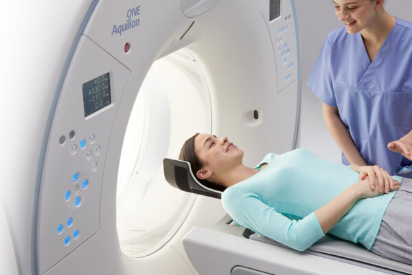
- Computed tomography (CT): builds cross-sectional images of bones, blood vessels and soft tissue. CT is crucial for finding cancers, cardiovascular disease and injuries.
- Magnetic resonance imaging (MRI): uses a magnetic field and radio waves to produce detailed images of organs and tissue. It is particularly good at brain, spinal cord and musculoskeletal problems.
- Mammography: a specialized X-ray of the breast that can find tumors too small to feel.
What early detection can look like
These two examples are illustrative, not real patients:
- Lung cancer. A 62-year-old former smoker who qualifies for screening has a low-dose CT. It shows a small nodule, follow-up imaging confirms an early-stage cancer, and surgery is curative.
- Heart disease. A 60-year-old with high blood pressure and mild chest discomfort has a CT angiogram, which shows a significant coronary blockage. Treatment opens the artery before a heart attack occurs.
Advances in imaging
Technology keeps making imaging more accurate and less invasive:
- Low-dose CT: reduces radiation while keeping image quality high, which makes CT suitable for screening people at high risk, such as long-term smokers.
- Contrast-enhanced imaging: contrast agents in MRI and CT make blood vessels and tissues easier to see.
- Digital and AI-assisted imaging: digital images are quick to share between providers, and AI tools can help radiologists spot subtle findings.
The importance of recommended screening
Following screening guidelines is one of the most effective ways to catch disease early. Examples include:
- Mammograms: recommended for women starting at age 40; ask your provider how often is right for you.
- Low-dose CT for lung cancer: recommended yearly for adults aged 50 to 80 with a 20 pack-year smoking history who smoke now or quit within the past 15 years.
- Bone density scans: recommended for women 65 and older, and for younger postmenopausal women at increased risk of osteoporosis.
Conclusion
Radiology is a cornerstone of modern medicine. It provides the insight that leads to early diagnosis and better outcomes, and imaging technology continues to improve.
Talk with your healthcare provider about the screening and imaging that fit your health needs. When you need an exam, our team in Laurel is here to help.
This article is general information and not medical advice. Talk with your healthcare provider about your own situation.